CERN Accelerating science

This website is no longer maintained. Its content may be obsolete. Please visit http://home.cern/ for current CERN information.

CERN Accelerating science

Synchrotron radiation imaging at 200 miles

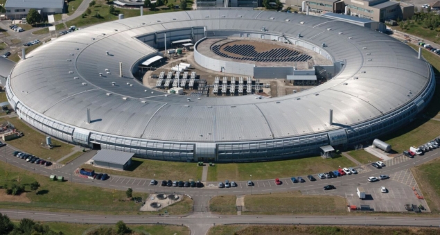
Whilst the world has been figuring out how to conduct business as usual remotely, experts from the University of Liverpool, based at the Cockcroft Institute, and Diamond Light Source, the UK’s national synchrotron in Oxfordshire, have taken this a step further and conducted a series of remote access beam measurements.

In this global lockdown period, a significant part of experimental physics has understandably been put on hold. With most of the world’s scientists unable to reach or access their experiments, efforts have shifted to simulations and planning for the future. A collaboration between the University of Liverpool and Diamond, which had installed an optical synchrotron radiation (OSR) imaging system in the 3 GeV ring at the synchrotron at the end of 2019, expected the same fate for their experiment.

However, as the OSR imaging system had been designed to be operated ~400m away in the Diamond control room; serendipitously this meant that thanks to some ingenuity with VPNs and video conferencing software, the experiment could be operated from ~200 miles away! With many of the synchrotron light sources around the world closed due to the pandemic, there was no guarantee that beamtime would be available at Diamond.

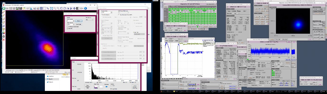
Fortuitously, thanks to the world-leading COVID-19 research being conducted at Diamond, the facility was still in operation. Several X-ray beamlines have been able to continue operation remotely throughout lockdown. These beamlines have been able to make significant contributions to the ongoing research into COVID-19, providing a deeper understanding of the virus, which could lead to the development of new therapies or treatments. The University of Liverpool’s measurements were completely non-invasive; they did not alter the beam at all for these ongoing important COVID-19 projects. Therefore, for the past few weeks a series of OSR experiments have been undertaken which aim to characterise the imaging capabilities of an existing diagnostic beamline, and hopefully shine a light on any beam halo effects. Fig. 1 shows the remote access screens. On the left experimental data from the Diamond tunnel as obtained by Liverpool experts is shown, whilst on the right various controls and beam parameters directly from the Diamond control room are shown.

Figure 1: Remote access and video conferencing being used for measurements at Diamond Light Source (Image: University of Liverpool/Diamond).

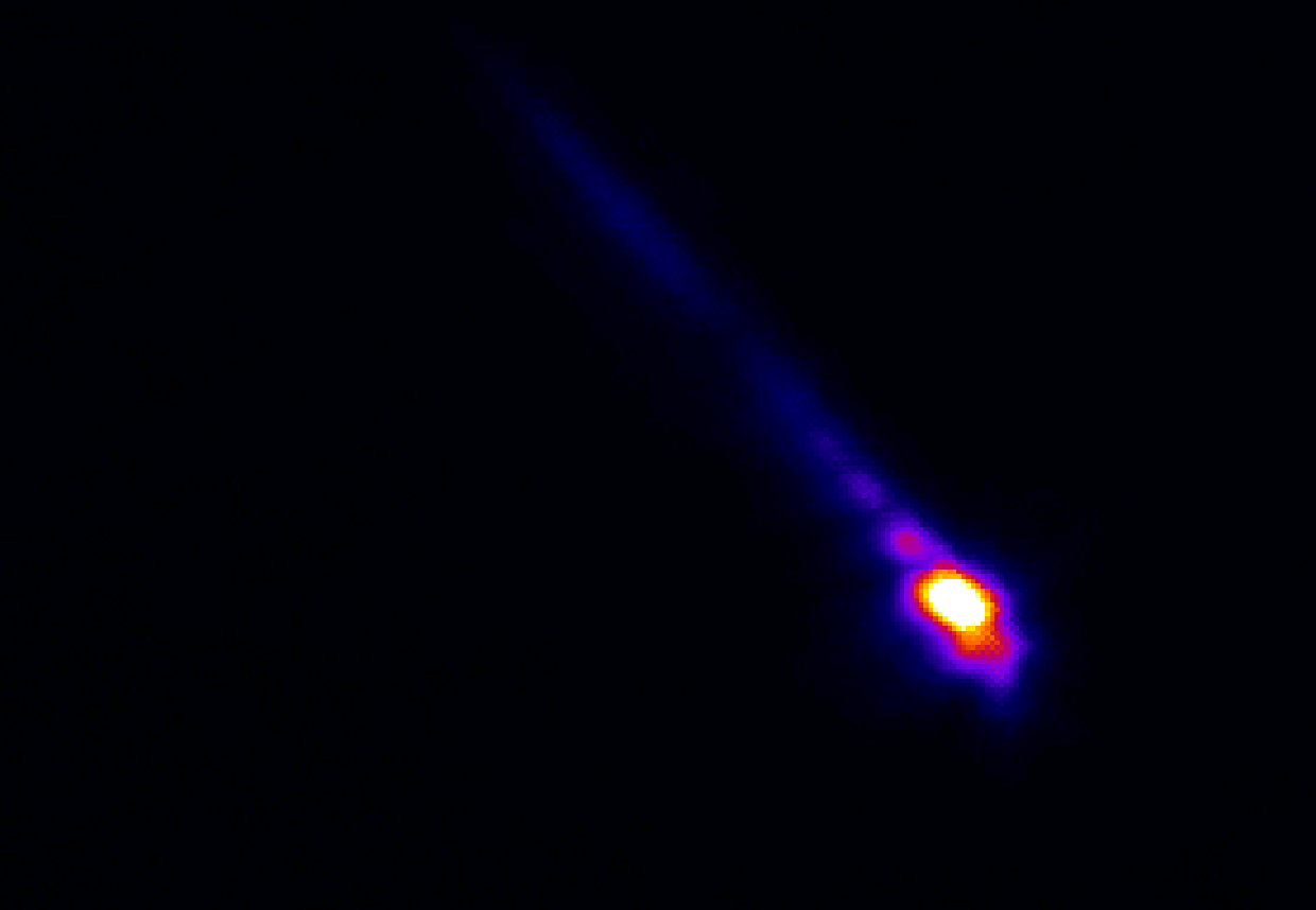
Beam halo is the relatively low intensity area of a beam which surrounds the core section. For high energy machines this low intensity is still an issue and can cause problems ranging from stored beam instabilities, all the way up to actual machine damage. The goal of beam halo studies is to observe the formation and extent of halo particles surrounding the beam core. In order to accomplish this it is important to understand the constituent parts of an OSR image; one such component is known as the “Filament Beam Spread Function” (FBSF), which is analogous to the “Point Spread Function” (PSF) found in classical optics. With a thorough understanding of the FBSF improved imaging techniques can be implemented, from Lyot stop imaging, to beam halo imaging. FBSF techniques can also be used to improve the typical resolution of OSR imaging methods.

Fig. 2 presents an example image of the 3 GeV electron beam at Diamond. The bright core is where the beam is located, and the elongated tail is the result of an accumulation of optical effects caused by various apertures along the beamline; these effects will be characterised in the FBSF. The group will now analyse the large quantity of data they have acquired. They will then use these results to guide improvements to the existing system and future measurements once the lockdown has passed.

Figure 2: 3 GeV Diamond beam image using optical synchrotron radiation (Image: University of Liverpool).

Dr Guenther Rehm, Head of Diamond’s Diagnostics group, commented:

“Despite the restrictions in place on movement and facility access, we were still keen on pursuing any possible avenue of our usual research efforts. Our ongoing collaboration with the University of Liverpool provided us with one such opportunity. Due to the manner in which the Liverpool installation was setup, remote operation was a prerequisite and measurements had no effect on the accelerator or its operation. By incorporating our ability to also control the accelerator operation remotely, we were able to perform a whole series of measurements with Liverpool without any member of the team being on the same site as the facility.

This period has forced all of us to think again on how we conduct our usual daily routine. In the case of this collaboration, we have been limited in what the measurements could achieve without physical access, but we have also produced a safe measurement program in the context of COVID-19. Alongside the usual beam-related implications of the data analysis, this effort to conduct research remotely and inclusively may shape the way we perform similar research in the future.”

Philippe Lebrun, JUAS Director
25th edition of Joint Universities Accelerator School
13 Mar 2018

25th edition of Joint Universities Accelerator School

Twenty-five years of training accelerator scientists and going from strength to strength

Athena Papageorgiou Koufidou, Livia Lapadatescu (CERN)
HIE-ISOLDE: challenges and future plans
15 Dec 2017

HIE-ISOLDE: challenges and future plans

HIE-ISOLDE advances the high energy frontier of the facility.

Panagiotis Charitos
Austrian synchrotron debuts carbon-ion cancer treatment
20 Oct 2019

Austrian synchrotron debuts carbon-ion cancer treatment

MedAustron, an advanced hadron-therapy centre in Austria, becomes one of six centres worldwide to treat tumours with carbon ions.

Synchrotrons on the frontline

Representation of the 3D structure of the main SARS-CoV-2 protease, obtained using Diamond Light Source. The coils represent “alpha” helices and the flatter arrows are “beta sheets”, with loops connecting them together. The organisation of alpha helices and beta sheets is often referred to as the secondary structure of the protein (with the primary sequence being the amino acid sequence and the tertiary structure being the overall 3D shape of the protein). (Image: D Owen/Diamond Light Source.)


At a time when many countries are locking down borders, limiting public gatherings, and encouraging isolation, the Diamond Light Source in Oxfordshire, UK, has been ramping up its intensity, albeit in an organised and controlled manner. The reason: these scientists are working tirelessly on drug-discovery efforts to quell COVID-19.

It is a story that requires fast detectors, reliable robotics and powerful computing infrastructures, artificial intelligence, and one of the brightest X-ray sources in the world. And it is made possible by international collaboration, dedication, determination and perseverance.

Synchrotron light sources are particle accelerators capable of producing incredibly bright X-rays, by forcing relativistic electrons to accelerate on curved trajectories. Around 50 facilities exist worldwide, enabling studies over a vast range of topics. Fanning out tangentially from Diamond’s 562-m circumference storage ring are more than 30 beamlines equipped with instrumentation to serve a multitude of user experiments. The intensely bright X-rays (corresponding to flux of around 9 × 10¹² photons per second) are necessary for determining the atomic structure of proteins, including the proteins which make up viruses. As such, synchrotron light sources around the world are interrupting their usual operations to work on mapping the structure of the SARS-CoV-2 virus.

Knowing the atomic structure of the virus is like knowing how the enemy thinks. A 3D visualisation of the building blocks of the structure at an atomic level would allow scientists to understand how the virus functions. Enzymes, the molecular machines that allow the virus to replicate, are key to this process. Scientists at Diamond are exploring the binding site of the main SARS-CoV-2 protease. A drug that binds to this enzyme’s active site would throw a chemical spanner in the works, blocking the virus’ ability to replicate and limiting the spread of the disease.

By way of reminder: Coronavirus is the family of viruses responsible for the common cold, MERS, SARS, etc. Novel coronavirus, aka SARS-CoV-2, is the newly discovered type of coronavirus, and COVID-19 is the disease which it causes.

Call to arms

On 26 January, Diamond’s life-sciences director, Dave Stuart, received a phone call from structural biologist Zihe Rao of ShanghaiTech University in China. Rao, along with his colleague Haitao Yang, had solved the structure of the main SARS-CoV-2 protease with a covalent inhibitor using the Shanghai Synchrotron Radiation Facility (SSRF) in China. Furthermore, they had made the solution freely and publicly available on the worldwide Protein Data Bank.

During the phone call, Rao informed Stuart that their work had been halted by a scheduled shutdown of the SSRF. The Diamond team rapidly mobilised. Since shipping biological samples from Shanghai at the height of the coronavirus in China was expected to be problematic, the team at Diamond ordered the synthetic gene. A synthetic gene can be generated provided the ordering of T, A, C and G nucleotides in the DNA sequence is known. That synthetic gene can be genetically engineered into a bacterium, in this case Escherichia. coli, which reads the sequence and generates the coronavirus protease in large enough quantities for the researchers at Diamond to determine its structure and screen for potential inhibitors.

Eleven days later on 10 February, the synthetic gene arrived. At this point, Martin Walsh, Diamond’s deputy director of life sciences, and his team (consisting of Claire Strain-Damerell, Petra Lukacik, and David Owen) dropped everything. With the gene in hand, the group immediately set up experimental trials to try to generate protein crystals. In order to determine the atomic structure, they needed a crystal containing millions of proteins in an ordered grid-like structure.

Diamond Light Source, the UK

Diamond Light Source, the UK’s national synchrotron facility, located at the Harwell Science and Innovation Campus in Oxfordshire. (Image: Diamond Light Source.)

X-ray radiation bright enough for the rapid analysis of protein structures can only be produced by a synchrotron light source. The X-rays are directed and focused down a beamline onto a crystal and, as they pass through it, they diffract. From the diffraction pattern, researchers can work backwards to determine the 3D electron density maps and the structure of the protein. The result is a complex curled ribbon-like structure with an intricate mess of twists and turns of the protein chain.

The Diamond team set up numerous trials trying to find the optimum conditions for crystallization of the SARS-CoV-2 protease to occur. They modified the pH, the precipitating compounds, chemical composition, protein to solution ratio… every parameter they could vary, they did. Every day they would produce a few thousand trials, of which only a few hundred would produce crystals, and even fewer would produce crystals of sufficient quality. Within a few days of receiving the gene, the first crystals were being produced. They were paltry and thin crystals but large enough to be tested on one of Diamond’s macromolecular crystallography beamlines.

Watching the results come through, Diamond postdoc David Owen described it as the first moment of intense excitement. With crystals that appeared to be “flat like a car wind shield,” he was dubious as to whether they would diffract at all. Nevertheless, the team placed the crystals in the beamline with a resignation that quickly turned into intense curiosity as the results started appearing before them. At that moment Owen remembers his doubts fading, as he thought, “this might just work!” And work it did. In fact, Owen recalls, “they diffracted beautifully.” These first diffraction patterns of the SARS-CoV-2 virus were recorded with a resolution of 1.9 Angstrom (1.9 × 10⁻¹⁰ m) — high enough resolution to see the position of all of the chemical groups that allow the protease to do its work.

By 19 February, through constant adjustments and learning, the team knew they could grow good-quality crystals quickly. It was time to bring in more colleagues. The XChem team at Diamond joined the mission to set up fragment-based screening – whereby a vast library of small molecules (“fragments”) are soaked into crystals of the viral protease. These fragments are significantly smaller and functionally simpler than most drug molecules and are a powerful approach to selecting candidates for early drug discovery. By 26 February, 600 crystals had been mounted and the first fragment screen launched. In parallel, the team had been making a series of sample to send to company in Oxford called Exscientia, which has set up an AI platform designed to expediate candidates in drug discovery.

Drug-discovery potential

As of early March, 1500 crystals and fragments have been analysed. Owen attributes the team’s success so far to the incredible amounts of data they could collect and analyse quickly. With huge numbers of data sets, they could pin down the parameters of the viral protease with a high degree of confidence. And with the synchrotron light source they were able to create and analyse the diffraction patterns rapidly. The same amount of data collected with a lab-based X-ray source would have taken approximately 10 years. At Diamond, they were able to collect the data in a few days of accumulated beamtime.

Synchrotron light sources all over the world have been granting priority and rapid access to researchers to support their efforts in discovering more about the virus. Researchers at the Advanced Photon Source in Argonne, US, and at Elettra Sincrotrone in Trieste, Italy are also trying to identify molecules effective against COVID-19, in an attempt to bring us closer to an effective vaccine or treatment. This week, the ESRF in Grenoble, France, announced that it will make its cryo-electron microscope facility available for use. The community has a platform called www.lightsources.org offering an overview of access and calls for proposals.

In addition to allowing the structure of tens of thousands of biological structures to be elucidated – such as that of the ribosome, which was recognised by the 2009 Nobel Prize in Chemistry — light sources have a strong pedigree in elucidating the structure of viruses. Development of common anti-viral medication that blocks the actions of virus in the body, such as Tamiflu or Relenza, also relied upon synchrotrons to reveal their atomic structure.

Mapping the SARS-CoV-2 protease structures bound to small chemical fragments, the Diamond team demonstrated a crystallography- and fragmentation-screen tour de force. The resulting and ongoing work is a crucial first step in developing a drug. Forgoing the usual academic root of peer-review, the Diamond team have made all of their results openly and freely available to help inform public heath response, limit the spread of the virus with the hope that this can fast-track effective treatment options.

This news first appeared on the CERN Courier

Alessandro Bertarelli (CERN)
High thermal performance materials
8 Oct 2018

High thermal performance materials

ARIES has launched an extensive characterization campaign of a broad range of high thermal performance materials for applications in future particle accelerators and the industry.

Marco Zanetti (INFN & Univ. Padua), Frank Zimmermann (CERN)
Workshop shines Light on Photon-Beam Interactions
7 Dec 2017

Workshop shines Light on Photon-Beam Interactions

The ARIES Photon Beams 2017 Workshop was held in Padua, Italy in late November 2017.

Ubaldo Iriso (ALBA-CELLS)
Different techniques of emittance measurements for SLS and FELs
2 Mar 2018

Different techniques of emittance measurements for SLS and FELs

The status of different techniques and some new approaches of emittance measurements for SLS and FELs were analyzed in a topical workshop at ALBA.

Different techniques of emittance measurements for SLS and FELs

The Topical Workshop on Emittance Measurements for Synchrotron Light Sources and FELs held at ALBA-CELLS in January 2018 brought together experts working on emittance measurements for electron machines, like Synchrotron Light Sources (SLS) and Free Electron Lasers (FEL). The workshop presented the status of the different techniques and discussed the challenges that this community is facing for the next generation of ultra-low emittance machines.

The emittance itself is not a direct measurable parameter and most accelerators monitor its surrogate: the electron beam size. In the case of SLS, the preferred techniques to infer the beam size are based on the analysis of the synchrotron radiation due to its non-destructive nature. In order to overcome the diffraction limit, direct imaging techniques like pinhole cameras or Compound Refractive Lenses use the x-ray part of the synchrotron radiation to avoid the diffraction limit. These techniques were reviewed and compared by L. Bobb (Diamond) and F. Ewald (ESRF), and it was concluded that they can be used down to the ~4µm level.

Other SLS like KEK or Max-IV use techniques based on the analysis of the synchrotron light coherence, like the double-slit interferometry presented by T. Mitsuhashi (KEK), or the polarization methods shown by A. Andersson (Max-IV). These techniques analyze the visible part of the synchrotron light, but in order to measure beam sizes below the ~2µm level, the system should be adapted to smaller wavelengths, like ultra-violet or even x-rays. This was the topic of A. Snigirev's (IKBF) presentation, highlighting the challenges of performing diffraction or interferometry in the x-rays part.

On the other hand, the beam sizes in FELs are measured through the interaction with obstacles in the electron beam trajectory, which often detrimentally affect the electron beam. Such is the case of Optical Transition Radiation (OTR) screens, which can be used for direct beam imaging or to produce Diffraction Radiation Interferometry, as shown by E. Chiadroni (INFN). Other obstacles like Wire Scanners (reviewed by K. Wittenburg from DESY) are nowadays getting thinner (down to the 1µm level) using lithography and electroplating, which improves the method resolution, thus allowing to measure beam sizes down to the 500nm level (S. Borrelli, SLS).  This level of resolution can also be achieved by using a laser wire, where the electron beam does not interact with a solid (metallic) object, but a “light pencil”, as shown by P. Karataek (JAI). However, this solution involves an important team of experts to maintain and operate the technique.

To measure the ultra-low emittance of the future SLS and FELs requires significant knowhow, while a new machine may need to combine different techniques, as presented by B. Yang (ANL) for the APS upgrade, whose design of the emittance measurement system already combines an x-ray pinhole camera, a diffractometer, and an x-ray interferometry. Perhaps this type of designs are the new path for the emittance measurement techniques. Finally, the workshop set forth new ideas like the beam size measurements using Heterodyne Speckle Fields or Cherenkov radiation, as well as a review of the methods used in other accelerators like hadron colliders or laser plasma accelerators.

The diagnostics experts on emittance measurements for SLS and FELs will strongly benefit from all the synergies between the different communities.


Header image: Group picture from the Topical Workshop on Emittance Measurements for SLS and FELs (Image credit: ALBA-CELLS)

Isabel Alonso, ‎Cedric Garion, Marco Morrone (CERN)
A new generation of beam screens
10 Dec 2018

A new generation of beam screens

The vacuum group of the HL-LHC collaboration had to innovate in a lot of aspects.

Daniela Antonio (CERN)
Building on the community’s collective experience on accelerator communication
25 Mar 2020

Building on the community’s collective experience on accelerator communication

Members from the accelerator communication community gathered at CERN for the 2nd Accelerator Communication and Outreach workshop.

Alexandra Welsch, Samantha Colosimo, Javier Resta López (University of Liverpool)
Accelerating Learning
8 Oct 2018

Accelerating Learning

Summer events held at CERN boost knowledge and collaboration. The events were coordinated by the QUASAR Group.