Cancer is a leading cause of death worldwide, with the disease accounting for an estimated 9.6 million deaths last year according to the World Health Organization. Although significant progress has been made in the use of proton and ion beams for cancer treatment, further R&D into the optimization of the underpinning accelerator technologies and advanced imaging systems is required to maximize the effectiveness of this technique.
This is the idea behind an international collaborative research network into the Optimization of Medical Accelerators (OMA). The project received 4 million € of funding from the EU within the Horizon 2020 programme to help improve ion beam therapy when it started in 2016; the network is now coming to an end and has achieved excellent results across its three closely interlinked work packages: Beam Imaging and Diagnostics, Treatment Optimization, as well as Facility Design and Optimization.
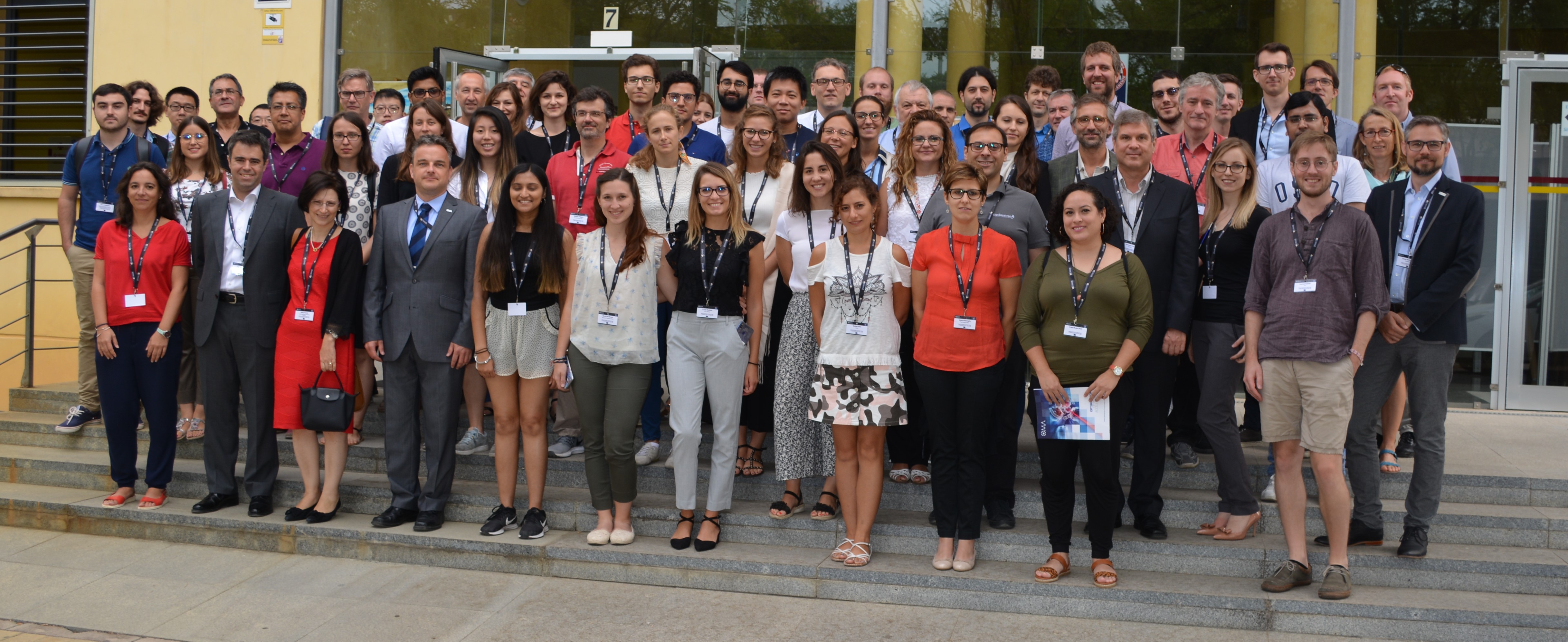
Group photograph during OMA conference in Seville, Spain. (Image: CNA)
The network has held its final official event in the form of an international conference on Medical Accelerators and Particle Therapy in Seville, Spain, 4-6 September 2019. More than 70 delegates joined at the headquarters of one of the project partners, the Centro Nacional de Aceleradores (CNA). A number of keynote talks covered for example next-generation therapy accelerators by IBA founder Yves Jongen, in-patient beam imaging by Katia Parodi/LMU Munich and dose delivery by Tony Lomax/PSI. The conference also offered a perfect environment for the OMA Fellows to present a summary of their research projects and findings.
OMA Fellow Sudharsan Srinivasan, who is based at PSI, showed results from R&D into a non-interceptive beam current and position monitor that uses the TM010 mode to characterize beams with currents down to 0.1 nA. His design combines mechanical simplicity with ease of manufacture due to its cylinder symmetry. Following characterization of the monitor on a test bench at PSI, Sud and co-workers carried out measurements with beam in the PROSCAN facility in summer 2019. They found very good signal linearity with beam position and low noise of the monitor, so that their monitor can now be considered for application in treatment beamlines.
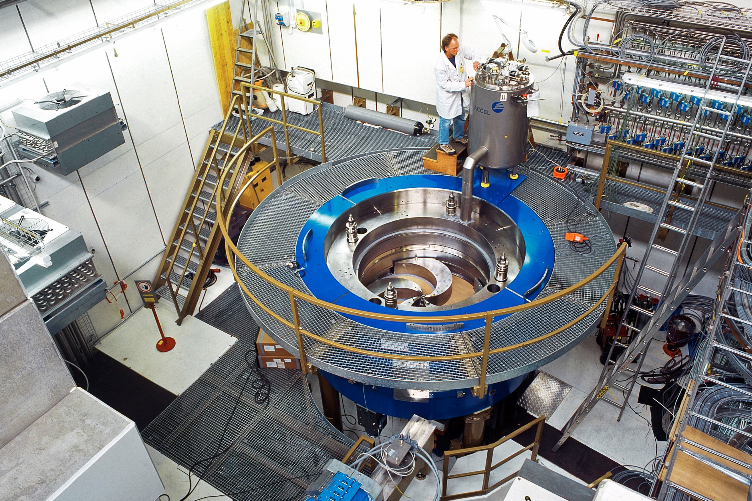
COMET cyclotron, part of the PROSCAN facility (Image: PSI)
Complementary to this development, Roland Schnuerer and OMA Fellow Jacinta Yap who are based at the University of Liverpool/Cockcroft Institute have investigated the use of the Vertex Locator (VELO) detector technology, originally developed for the Large Hadron Collider beauty experiment (LHCb) as a non-interceptive online beam monitor. Due to the semi-circular cut-out in the sensor, a precise measurement of the beam halo without interfering with the beam core is made possible. By correlating the beam halo reading with data from a Faraday Cup, a halo-dose correlation data base for different settings can be established and used for QA purposes. Proof-of-principle measurements using this monitor at the 40 MeV proton line at the University of Birmingham were completed earlier in 2019 and demonstrated VELO’s unique proton counting capability. Supported by detailed beam tracking studies on the example of the Clatterbridge Cancer Centre, the researchers have shown how VELO can be used for least invasive online beam monitoring and QA.
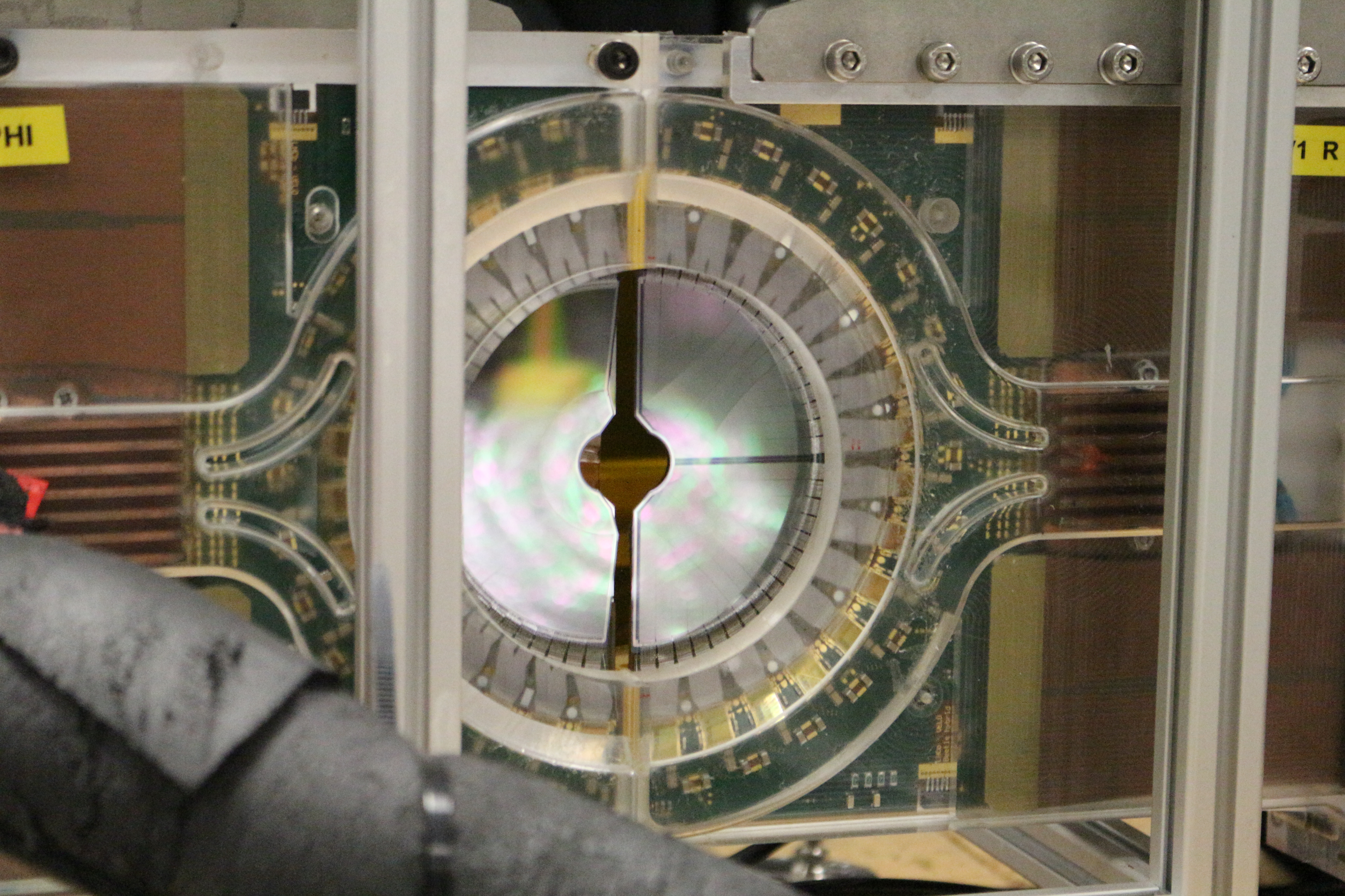
VELO detector (Image: University of Liverpool)
In addition to novel beam monitoring techniques, OMA Fellows also carried out research into 3D/4D-patient imaging. These are used to position the patient before the therapy and also to monitor the patient position during treatment so that moving tumors can be treated and that the beam can be stopped if the patient is in the wrong position. Samuele Cotta who was based at German company ViALUX focuses on studies into errors and potential damage of the scanners from secondary radiation. Using the intra-network links within OMA, he carried out measurements at several facilities including GSI’s carbon ion treatment room. His studies showed only few disconnections when the monitor was subject to a 1,000 h accelerated test and allowed improving the sensor by implementing a fast automatic hardware reset in the case of a disconnect event. Further long-term tests are planned in a radiotherapy environment to validate scanner radiation hardness under real treatment conditions.
OMA Fellow Liheng Tian who is based at LMU in Germany presented new treatment planning approaches accounting for in-vivo proton range verification. Based on previous work at his host to re-optimize treatment plans using spot-by-spot conformities of prompt gammas and dose, an improved approach was studied and he showed how this can help ensure enhanced robustness of prompt gamma-dose correlation of boosted pencil beams in the presence of interfractional anatomical changes. In his talk, he also discussed potential advantages over other proposed methods, such as spot aggregation.
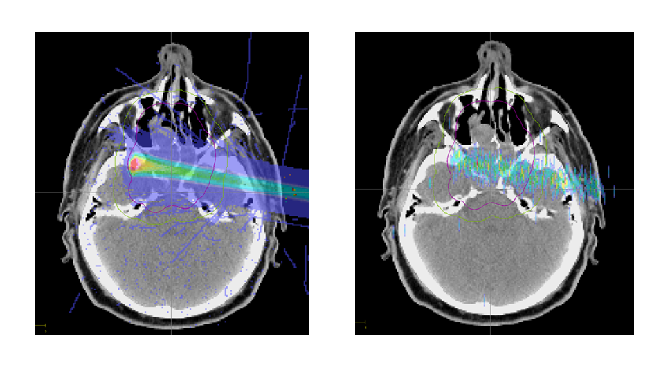
Left: Dose distribution of a given pencil beam. Right: Prompt gamma emission distribution of this pencil beam. (Images: LMU)
Professor Welsch who initiated and coordinates the project said: “OMA has been a fascinating journey for all of us. It makes me incredibly proud to see the excellent research results of our Fellows here at this conference. The Fellows have followed a unique training programme, which I am sure will provide them with an excellent basis for their future careers.”
The OMA Fellows have received extensive research-based training within a truly unique international partnership. The fundamental core of their training consisted of dedicated cutting-edge research projects for each Fellow, combined with an intra-network secondment scheme. This training concept is based on the previous DITANET, oPAC and LA3NET MSCA networks and is expected to serve as an excellent basis for future initiatives. In addition, OMA has organized a number of international schools and topical workshops, which were open to the wider community. These events were so popular that the network has decided to continue them beyond the lifetime of the project. This way, OMA will continue to help build bridges between the accelerator and medical community, contributing to a further optimization of ion beam therapy.


